Mouth cancer
Contents
By far the most common form of oral cancer is squamous cell carcinoma (SCC). SCC is a malignancy of the cells lining the lips, mouth and throat. These are cells similar to those of skin but don’t under normal circumstances have the thicker layer of keratin that skin has.
Causes
Most oral cancer (as in most of upper aerodigestive tract cancer) is due to tobacco and alcohol consumption to “excess” (over 75%). Our knowledge of risk factors associated with oral cancer is principally that of squamous cell carcinoma. There is relatively little known about the aetiology of salivary cancer, sarcoma or mucosal melanoma.
Tobacco
There is overwhelming evidence that tobacco usage in the form of cigarette, cigar and pipe smoking is associated with a more than 10-fold increase of the risk of developing oral cancer in comparison to non-users. All forms of tobacco smoking are associated with increased risk. There is also evidence that heavier and prolonged usage of tobacco is associated with a greater risk of having oral cancer as well as a second malignancy. Smoking cessation is worthwhile as the risk after abstention for more than 10 years becomes similar to that of non-smokers.
The mechanism of tobacco carcinogenesis has been well researched. There are several hundred carcinogens identified in tobacco smoke, e.g. aromatic hydrocarbons and tobacco specific nitrogen compounds. These agents act both locally and systemically. They interfere with DNA replication leading to mutations, and contribute to the molecular chain of events in malignant transformation.
Chewing tobacco with or without areca (betel) nut and snuff dipping (placing it buccally) are both associated with oral cancer The areca nut is itself an oral carcinogen. It is associated with submucous fibrosis and epithelial dysplasia, leading on to malignant transformation particularly if used in conjunction with tobacco. The areca nut is chewed as a quid of betel leaves and slaked lime, with or without tobacco. It is popular in the Indian sub-continent, South East Asia and New Guinea.
Alcohol consumption
As there is a tendency for those who smoke to also consume alcohol, often in significant quantities, care is needed in interpreting their association with oral cancer. There is good evidence that heavy alcohol consumption is associated with a significant risk of oral and pharyngeal cancer. For example, consuming 100g of alcohol or more per day increases the risk of developing oral cancer by at least six-fold. Although certain alcoholic beverages may have significant contributory impurities, it is thought that the quantity of alcohol consumed is the main significant risk factor.
Alcohol is thought to contribute to cancer of the head and neck by local and systemic mechanisms. These include ethanol-induced increased permeability of oral mucosa (to carcinogens), active metabolites of ethanol being carcinogenic, heavy drinkers with liver disease resulting in reduced detoxification of carcinogens and poor diet associated with alcoholics.
Synergistic effect of smoking and alcohol
Smoking and high alcohol consumption is associated with a synergistic or multiplicative effect in terms of risk of oral cancer. Heavy drinkers who also smoke a lot have an over 35 times greater risk in comparison to those who do not smoke or drink.
As some 75% of those with oral cancer smoke and/or drink alcohol, these factors should be targeted in preventing oral cancer.
Other factors
Consumption of fruit and vegetables has a protective beneficial effect. Certain diets such as salted fish (Chinese) had been reported to have an increased risk of pharyngeal cancer.
Certain viruses are known to be closely related to human cancer. For example, human immunodeficiency virus (HIV) with Kaposi’s sarcoma, Epstein Barr virus in nasopharyngeal carcinoma, Hepatitis B virus in hepatocellular carcinoma and Human Papilloma Virus (HPV) in cervical and anogenital cancer. The HPV/oral cancer link is currently being investigated. The HPV 16 and 18 strains are particularly implicated, and are associated with base of tongue/tonsillar cancers, which has seen a marked increase in recent years (up to 15% per annum). People affected tend to be younger, are less likely to be smokers, and (if non-smokers) have much better outcomes in terms of survival. This has led to a number of studies involving treatment intensity reduction in those treated by radiotherapy in a bid to reduce the very severe morbidity associated with many of the more recent non-surgical oncological treatments (such as radiotherapy and particularly chemoradiotherapy).
Irradiated tissue has a long-term risk of forming new malignancies. This is one of the many reasons to look at non-radiation options in treating cancers in younger patients.
Poor dental status had often been quoted as a cause of oral cancer. However, these patients often have confounding factors such as poor socio-economic status, heavy alcohol and tobacco usage as well as poor dietary habits.
Premalignant conditions/lesions
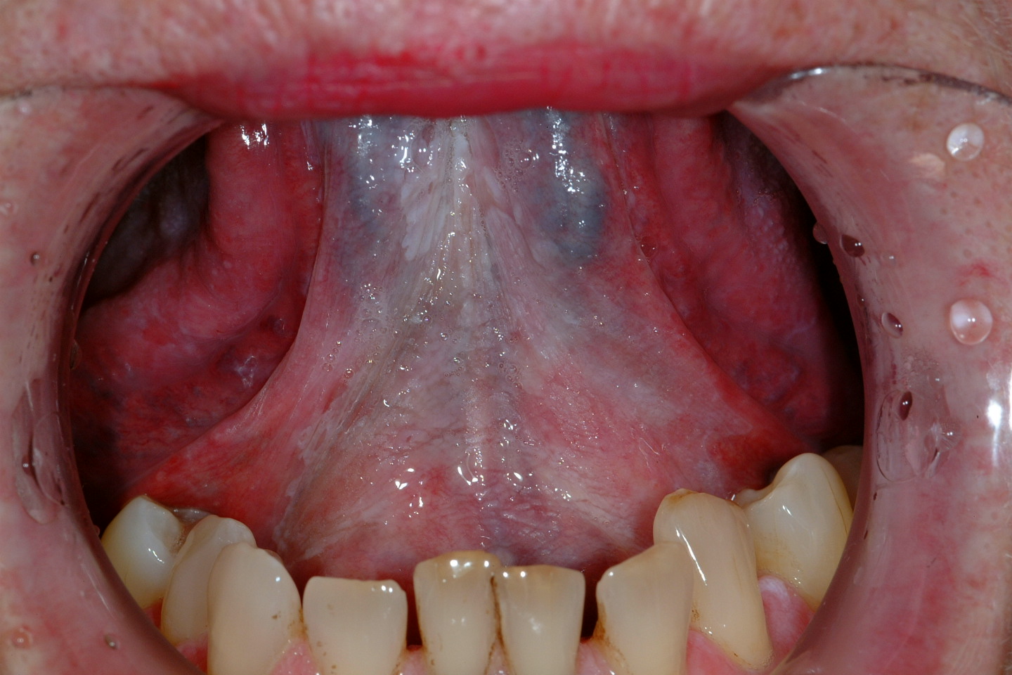
An oral leukoplakia (WHO definition) is a white patch or plaque that cannot be characterized, clinically or histopathologically, as any other disease. Erythroplakia can be similarly described but for bright red patches or plaques. Erythroleukoplakia (speckled leukoplakia) forms the third lesion commonly associated with risk of malignant transformation. This risk has been reported to be 2-6% for leukoplakia (varies with sites, e.g. highest in floor of mouth). 40-50% of erythroplakias have high grade dysplasia or carcinoma present. Speckled leukoplakia is of intermediate risk.
Other lesions associated with malignant transformation include chronic hyperplastic candidiasis, submucous fibrosis and sideropenic dysphagia. Less commonly, certain conditions such as dyskeratosis congenita (tongue leukoplakia); discoid lupus erythematosus (lip cancer) and lichen planus (1-3% incidence, lichenoid dysplasia main reason) have a risk of malignant change.
The management of these premalignant lesions, in particular, leukoplakia, can be controversial. Cessation of smoking is beneficial, with spontaneous resolution seen in some 50% of lesions in some studies. Some clinicians prefer to closely observe these lesions with selective biopsies where there are worrying changes such as induration, contact bleeding, ulceration or conversion to erythroplakia. Surgeons, in particular, may advocate laser excision with the aim of achieving thorough histological analysis and reducing the incidence of malignant transformation.
Decision making depends on several issues such as whether the lesions are localized (easy to remove) or widespread, patient compliance and preference as well as clinician’s resources. Biopsies are often carried out to assess the degree of dysplasia, in particular where there is speckled leukoplakia or erythroplakia. A large proportion of these lesions have high grade dysplasia if not malignant transformation.
Vitamin A and related compounds (retinoids) have been used as chemo-preventive agents. Some studies have demonstrated the resolution of leukoplakia with regular usage of retinoids. However, the side-effects associated with retinoids usage can be considerable. Poor compliance and recurrence of lesions on cessation of these chemo-preventive agents have limited the usefulness of this treatment modality.
The use of NSAIDs in chemoprevention of oral cancer is unsupported by evidence and is a good example of research fraud.
Diagnosis journey
Presentation
Oral cancer can be asymptomatic for some time, resulting in late presentation. In addition, a significant proportion of oral cancer sufferers are alcohol dependent recluses.
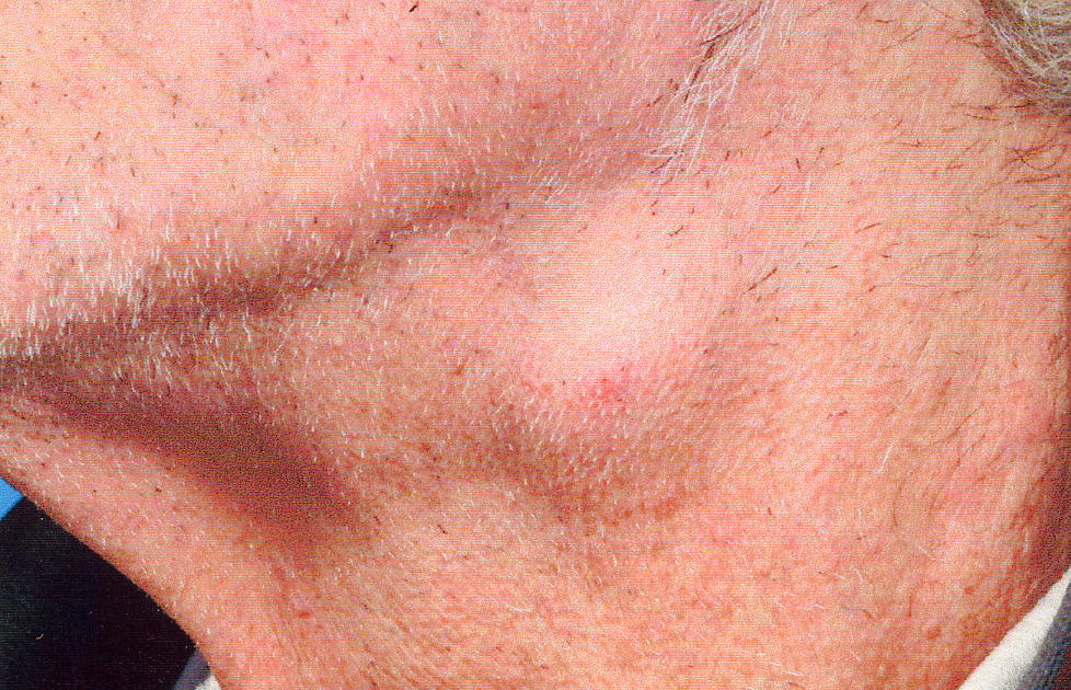
Early symptoms and signs of oral cancer include persistent mouth ulceration (often painless), lumps, white, red or speckled lesions. Later, features such as recent onset of difficulty with speech or swallowing, numbness, neck swelling (lymphadenopathy) and unexplained loosening of tooth/teeth. Any of these persistent lesions (more than 3 weeks) should be referred urgently for specialist opinion and possible biopsy. Although the vast majority of oral cancer is clinically apparent, both patients and general practitioners may miss a small proportion. Some can present late with enlarging neck nodes.
Fine needle aspiration cytology examination (using a needle and syringe to aspirate cells from a lump in an outpatient clinic setting) can be useful diagnostically.
The oral mucosa has a renowned ability to heal. It is therefore important for the specialist to see the suspected lesion intact, i.e. leave the biopsy procedure to them. This will enable better recording and more accurate treatment planning.
Techniques to reveal high risk lesions are available, e.g. Toluidine blue rinse, optical techniques. It is generally accepted that this method is not suitable for general use due to a significant proportion of false positive and negative results.
There is a drive from both the professionals and the government to improve outcome in head and neck cancer. In the state controlled systems of the UK this has produced a top down dogmatic administrative approach which has mixed benefits. These efforts included setting standards in quality and quantitative parameters in the form of guidelines, recommendations, political cancer strategies by various governments and expert group consensus views (in the UK NICE produced a document on head and neck cancer).
Data collection, referral pattern, fast track system, delays
Data collection for cancer statistics is vital for improving outcome. In the UK the HANA project aims to standardize this aspect and enable auditing and research on a national basis. The UK government Cancer Strategy target for urgent referrals is aimed at reducing delays in urgent referral to appropriate cancer specialists. There is however the real risk of delays for cancer cases that had not been referred urgently. Part of these political initiatives in the UK, are driven by unfavourable comparisons in cancer survival (in general) between the UK and other developed European countries. There is a degree of frustration with nationwide broad brushstroke approaches to “national cancer statistics” as at least one pan-European survey showed Scotland to have one of the best 5-year survival rates for oral cancer when analysed specifically. “Lies, damned lies and statistics” is a cliché for a reason.
The diagnosis of mouth cancer is a major life event for the patient. It is important to ensure that a histological diagnosis is obtained as even experienced specialists can make a wrong diagnosis based on clinical grounds alone.
Dealing with bad news
This should be carried out in a sympathetic, unhurried manner within a quiet and private environment. Patients on receiving the news can become significantly stressed psychologically and not able to take in or retain much information. It is important that support is available such as a trained nurse counselor, a contact number to ring and written information with or without an information leaflet on oral cancer (that the patient can read). The general medical practitioner, family physician or other referral source should be informed promptly.
Diagnostic work up
In addition to history and clinical examination, further investigations are necessary to evaluate the extent of the disease and the fitness of the patient to undergo treatment both medically and psycho-socially.
Assessment in practice
The key questions to answer are tumour resectability (and proximity to vital structures), likelihood of neck (both sides) involvement and presence/absence of distant metastases.
Primary Tumour
Clinical assessment often yields a lot of information on its own within the oral cavity. However, occasionally, other methods are required to assess e.g. extension of tumour into pharynx, closeness to mandible, or retching or pain excluding a thorough examination. MRI is better than CT as the latter are often degraded by associated amalgam artifacts. More information on bone invasion can be gained using plain radiographs, MRI or CT scan and very rarely radio-isotope scintigraphic bone scans.
Neck metastases
Clinical palpation tends to miss some 30-40% of neck metastases. Imaging techniques such as ultrasound, MRI or CT scanning and ultrasound-guided fine needle aspiration provides better accuracy (in ascending order). If a thorough histological analysis of neck specimens in clinically and radiologically negative necks is carried out some 30% of oral cancer patients will have occult disease in the neck.
Distant metastases
The vast majority of distant metastases arising from oral SCC occur in the thorax. CT of the thorax is more detailed and accurate than plain chest radiography and is usual in UK practice.
Staging & treatment plan
Staging of the disease
It is important that the staging nomenclature adopted is widely accepted and therefore must be simple and clearly defined. The staging process allows the clinician to have a uniformly communicable description of that patient’s disease, i.e. the extent of the cancer. It helps in the treatment plan process, predicts prognosis and enables better audit (within and between different centres) as well as meaningful multi-centre studies.
Tables 1 and 2 show the current staging nomenclature in use for oral squamous cell carcinoma (SCC).
| T – Primary Tumour | |
|---|---|
| TX | primary tumour cannot be assessed |
| T0 | No evidence of primary tumour |
| TCIS | carcinoma in situ |
| T1 | Tumour 2 cm or less in maximum dimension |
| T3 | Tumour more than 4 cm in maximum dimension |
| T4 | Tumour invades adjacent structures, e.g. bone, deep extrinsic muscles, maxillary sinus or skin |
| N - Regional lymph nodes | |
| NX | regional lymph node s cannot be assessed |
| N0 | No regional lymph node metastasis |
| N1 | metastasis in a single ipsilateral lymph node 3 cm or less in maximum dimension |
| N2a | metastasis in a single ipsilateral lymph node more than 3 cm but not more than 6 cm maximum dimension |
| N2b |
metastases in multiple ipsilateral lymph nodes none more than 6 cm maximum dimension |
| N2c |
metastasis in contralateral or bilateral lymph nodes none more than 6 cm maximum dimension |
| N3 |
metastasis in a lymph node more than 6 cm maximum dimension |
| M – Distant metastases | |
| MX | presence of distant metastasis cannot be asessed |
| M0 | no distant metastasis |
| M1 | presence of fistant metastasis |
| Stage | T status | N status | M status |
|---|---|---|---|
|
Stage 0 |
TCIS |
N0 |
M0 |
|
Stage I |
T1 |
N0 |
M0 |
|
Stage II |
T2 |
N0 |
M0 |
|
Stage III |
T3 T1, T2, T3 |
N0 N1 |
M0 M0 |
|
Stage IVA |
T4 T4 T1, T2, T3, T4 |
N0 N1 N1, N2 |
M0 M0 M0 |
|
Stage IVB |
T1, T2, T3, T4 |
N3 |
M0 |
|
Stage IVC |
T1, T2, T3, T4 |
N0, N1, N2, N3 |
M1 |
There are other staging methods. However, to be effective, a staging method needs to be widely known (and therefore adopted universally), easy to use, reliable to score and ideally able to fulfill its roles stated above. Despite its several inadequacies the TNM system remains the most universally used.
Once the staging process is completed, the definitive treatment plan can be formed. Treatment planning is generally carried out in a multi-disciplinary team setting. The team usually consists of head and neck cancer specialists; surgeon(s), clinical oncologist(s) with radiotherapy expertise, pathologist, radiologist as well as allied medical professions such as speech and language therapist, dietician, head and neck cancer nurse specialist. There are also other specialists associated with the service such as dental hygienist, maxillofacial technician and occupational therapist, psychologist and physiotherapist. The team approach is aimed at removing bias in treatment planning and outcome assessment and combining experience as well as expertise to ensure the best possible treatment can be provided for the patient.
Treatment plan
It is important to be clear as to the treatment intention for that individual patient. In the majority of cases, this is curative, in some cases it is entirely palliative which may be defined as “active” (usually using low dose low toxicity radiotherapy or chemotherapy) or best supportive care. Rarely “curative treatment with palliative intent” that is the same intensity of treatment; often surgery and radiotherapy is offered in the belief that even if the cancer is not curable on the grounds of metastases the best chance of effective symptom control lies with curative intensity treatment.
Treatment intent
The overall decision on accepting treatment must lie with the patient (when a competent adult). This will be made by balancing the information on acute and chronic morbidity of treatment, relative chances of cure, morbidity of not having optimum treatment and individual quality of life decisions. All patients are different and each will have differing priorities, they should be enabled to make the most appropriate decision for them, ideally with an adequate length of time, support and environment to come to a balanced decision.
Over 90% of mouth cancer is squamous cell carcinoma. The natural progression of the disease involves invasion of local structures, spread to the regional cervical nodes and distant sites, principally the lungs. The invasive process is such that the microscopic front tends to be further than macroscopically apparent. The more advanced the disease, the poorer the prognosis.