Cleft lip/palate
Cleft lip and palate
Clefts of the lip and palate are the most common craniofacial malformations, and comprise 65 % of all anomalies affecting the head and neck. There are two useful distinct types of cleft anomaly, cleft lip with or without cleft palate, and isolated cleft palate. There is a difference in the pathogenesis of the two.
Aetiology
The aetiology of clefting is multi-factorial. Some environmental factors such as phenytoin (an anti-epileptic drug) taken in pregnancy increase the risk of cleft lip and palate, other drugs such as retinoids (used to treat a range of skin conditions), folate deficiency (increased need during pregnancy) or the foetal alcohol syndrome also increase the incidence. Maternal obesity increases the risk. Folic acid supplementation has been shown to reduce the incidence.
The term for this overall failure of normal development is that the formation of cleft lip and palate exhibits polygenic inheritance with a threshold.
Defects caused by the clefting process
The disruption of the muscular sphincter of the mouth (orbicularis oris) may be partial, as in an incomplete cleft of the lip (see Figure 1), or total as in a complete unilateral cleft of the lip (see Figure 2).
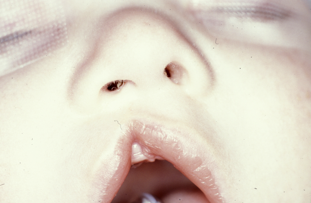
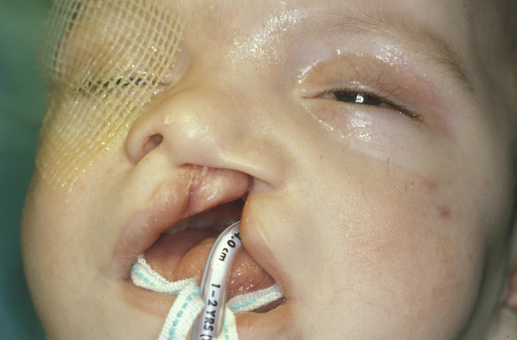
This has a direct effect on the associated deformity. Patients with incomplete cleft lips often seem to have relatively normal alar bases and intra alar widths (wings and width of the nose). Patients with complete clefts will have a significant lateral and posterior migration of the alar base on the cleft side. A bulge around the region of the alar base will be seen where the abnormal insertion of the orbicularis oris muscles are and a bulge lateral to the alar base where the external nasal muscles are attached is seen. In complete clefts there will often be a defect in the alveolus (socket in the jaw bone to hold teeth) which may range from a slight notch to a complete cleft of the alveolus extending into the palate. In complete clefts of the lip, the nasal septum and collumella (skin and cartilage separating the nostrils) are pulled to the side opposite the cleft by the contralateral normally attached musculature. This leads to a deviation of the tip of the nose and contributes to the classical appearance.
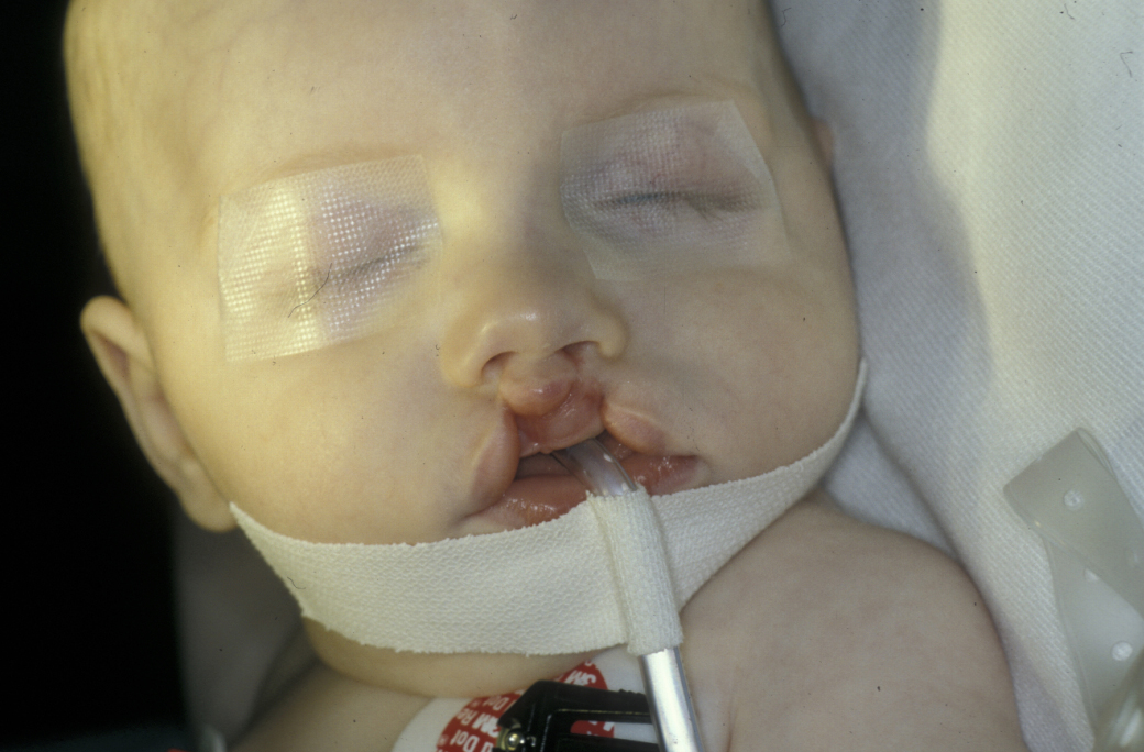
Patients with bilateral clefts may exhibit more apparent symmetry in both incomplete (see Figure 3) and complete cases (see Fig 4) but the main problem is that the premaxilla (bones at the tip of the upper jaw) will rotate forward as it contains no muscle and the orbicularis oris muscle exerts no restraints on the forward growth of the premaxilla.

In clefts of the alveolus whether bilateral or unilateral, the defect in the alveolus may be either simple notching or notching associated with congenital absence of the lateral incisor (second front tooth) or presence of supernumeraries (extra teeth), or a rudimentary lateral incisor, or a complete oro-nasal fistula with a range of associated dental abnormalities.
If the defect extends between the lip and the palate, any combination of complete or incomplete, bilateral or unilateral clefts of the lip and alveolus may be associated with the cleft extending through the hard and soft palate. In unilateral cases, rotation and collapse of the segments of the maxilla (upper jaw) in an inward and anterior (forward) direction is usually seen. This is more marked on the side of the cleft. This part of the cleft maxilla is referred to as the lesser segment. In bilateral clefts, both the lateral segments are collapsed behind the prominent premaxilla.
Isolated clefts of the palate may affect the soft palate or the hard palate, or a combination of both, or the uvula (soft tissue structure at the back of the soft palate) in isolation. There is also a condition known as submucous clefting where the oral mucosa (lining) of the palate, and often the oral and nasal mucosa of the palate are intact, but there is no underlying bone or muscle. This condition can be easily missed and may only become obvious once the child has had its adenoids (tonsils) removed and develops marked hyponasality in speech. The usual finding is a widening of the dental arch posterioraly (to the back), associated with the cleft and significant eustachian tube (structure connecting the middle ear to the part of the throat behind the nose) dysfunction which frequently results in otitis media (glue ear) and hearing impairment.
The common marks of cleft lip and palate include problems with feeding due to a communication between the mouth and the nasal cavity, decreasing the baby’s ability to suck. This can be overcome by positional feeding and the use of appropriate bottles to deliver feed.
Problems with hearing
Eustachian tube dysfunction as a result of cleft palate inhibits adequate ventilation of the middle ear and this causes secretory otitis media (glue ear) and can result in deafness. This contributes to a child’s difficulty in learning speech as hearing oneself talk is part of the natural bio-feedback of learning to communicate.
Direct speech problems
Learning to speak with lip or alveolar defects, defects in the hard and soft palate, and in later life dental anomalies have an obvious impact and require early assessment and, if necessary, intervention by speech and language therapists.
The anatomical problems related to the nose can result in a poor nasal airway with resulting speech, breathing and sinus related problems.
Principles of management
In many centres, neonatal detection by ultrasound scans is possible as the shape of the face is detectable by the 14th week in utero and the parents may well be advised that their child will have a cleft. This may help to come to terms with the shock of having a child with a cleft. It is however common for parents to experience feelings of guilt and regret, and often requires time to effectively grieve for the loss of the anticipated ‘normal’ child. It is sometimes helpful to take photographs of the child with the cleft as this child effectively vanishes once the initial cleft repair has been carried out.
At the point of diagnosis, home support by a specialist health visitor to assist with bonding between parents and child and help with feeding can be useful. Directed flow of milk into the mouth using soft bottles and positioning the child will allow the child to be adequately nourished. A range of useful bottles and teats are available and can be obtained in the UK from the Cleft Lip and Palate Association (CLAPA), a cleft support group.
Most centres in the UK have limited active intervention at this early time although world-wide a variety of pre-surgical orthopaedic techniques have been advocated at this point. Although practice varies throughout the world these are not currently in vogue in the UK.
Timing of surgery
The precise details of the timing of surgery will vary depending on the individual. A commonly used protocol includes:
- lip +/- alveolus +/- soft palate repair at three to six months old;
- repair of residual palatal defect at nine to twelve months old;
- audiology (hearing assessment) +/- myringotomy Intervention to release pressure on middle ear) either at this time or as indicated by audiological assessment;
- formal speech assessment around two-years old;
- pharyngeal (throat) surgery if indicated by speech and language therapy assessment at four to five-years old;
- alveolar bone grafting assessment at age eight years, followed by actual alveolar bone grafting between the ages of nine and eleven;
- definitive orthodontics (treatment of irregularities of teeth) +/- orthognathic surgery (corrective jaw surgery) between the ages of 15 and 18 years old;
- definitive rhinoplasty (corrective nose surgery) plus definitive restorative dentistry age 18 years plus.
Primary dentition and speech assessment
The development of the primary dentition should be allowed to progress essentially naturally with good paediatric dentistry in order to maintain space. No orthodontic intervention in the primary dentition is warranted, good preventive dentistry is. A joint ENT (ear, nose & throat) and speech therapy assessment should be carried out to detect middle ear effusions and appropriate development of speech patterns. Hypernasal speech and/or glue ear should be addressed, either by therapy or surgery such as pharyngoplasty or myringotomy (for glue ear).
Mixed dentition
At this stage the growth retarding effects of initial surgery become more evident, this is predominately side to side in the top jaw, and then front to back as growth in this dimension predominates with age. The eruption of the permanent incisors demonstrates defects in enamel formation and position, and the upper incisors may erupt into lingual occlusion. Orthodontic preparation for alveolar bone grafting can usefully be combined with limited alignment of the incisors in the mixed dentition.
Alveolar bone grafting
This technique provides bone through which a permanent canine and lateral incisor can erupt into the arch which unifies the maxilla, providing a stable intact arch, improve the alar base support, closes the residual oro-nasal fistula and provides a palatal base for later orthognathic surgery.
Permanent dentition
The establishment of the permanent dentition including guiding of the canines, which have to be exposed in about 15 % of cases, will basically be a decision as to whether or not orthodontic camouflage or orthognathic surgery is indicated.
Rhinoplasty
Rhinoplasty should be performed at the end of treatment rather than as an intermediary as the final position of the nose can only be predicted once the maxilla is in a stable position.
Definitive restorative dentistry
Implant-borne permanent prosthesis, or fixed-bridge prosthesis which can allow optimal oral hygiene should allow full and final rehabilitation of cleft patients.