Jaw joint problems
Contents
Temporomandibular joint pain dysfunction syndrome (TMJPDS)
This is a descriptive term (which often means very little is known about it) relating to pain arising from the joint itself and the muscles of mastication. It is also known as facial arthromyalgia and myofacial pain.
It has been suggested that up to 70% of the population have, at some time, experienced at least one episode of painful symptoms that can be attributed to TMJ dysfunction.
The aetiology of TMJPDS is unknown, although stress, often associated with ‘parafunctional habits’ such as bruxism (teeth clenching) are common features. Headaches, neck ache and back pains are also commonly seen with this condition. Abnormalities in occlusion have long been held responsible for TMJPDS, but this is controversial and the evidence for this is conflicting. There does appear to be a subgroup of people who do benefit from correction of a malocclusion; however this is certainly not the case in all people. Furthermore, it is important to identify TMJPDS in people who are about to undergo orthognathic surgery (corrective jaw surgery) as in a proportion of these, surgery or even orthodontics will make things worse.
Three cardinal features exist:
- pain
- joint noise
- restricted movement
Pain is the most common complaint and is by far the most difficult component to assess. It is characterised clinically by pain in and around the jaw joint, involving the muscles of mastication, and often radiating to the temple, jaw and into the neck. Patients often describe a dull, deep ache around the joint, which may resemble ‘earache’, along with a superimposed sharper component, which may radiate over the side of the face. Clicking of the joints with limitation of the range of mouth opening is also commonly reported. Joint noises, however, are quite common in asymptomatic people in the general population. Joint noises are of little clinical importance in the absence of pain. When mechanical symptoms predominate, the term ‘internal derangement’ (see below) is sometimes used, implying that there is a problem with the meniscus. However clicking is a common finding in the normal population and in the absence of other symptoms does not need treatment.
Clinical examination includes assessment for tenderness in the muscles of mastication (see Figure 1).
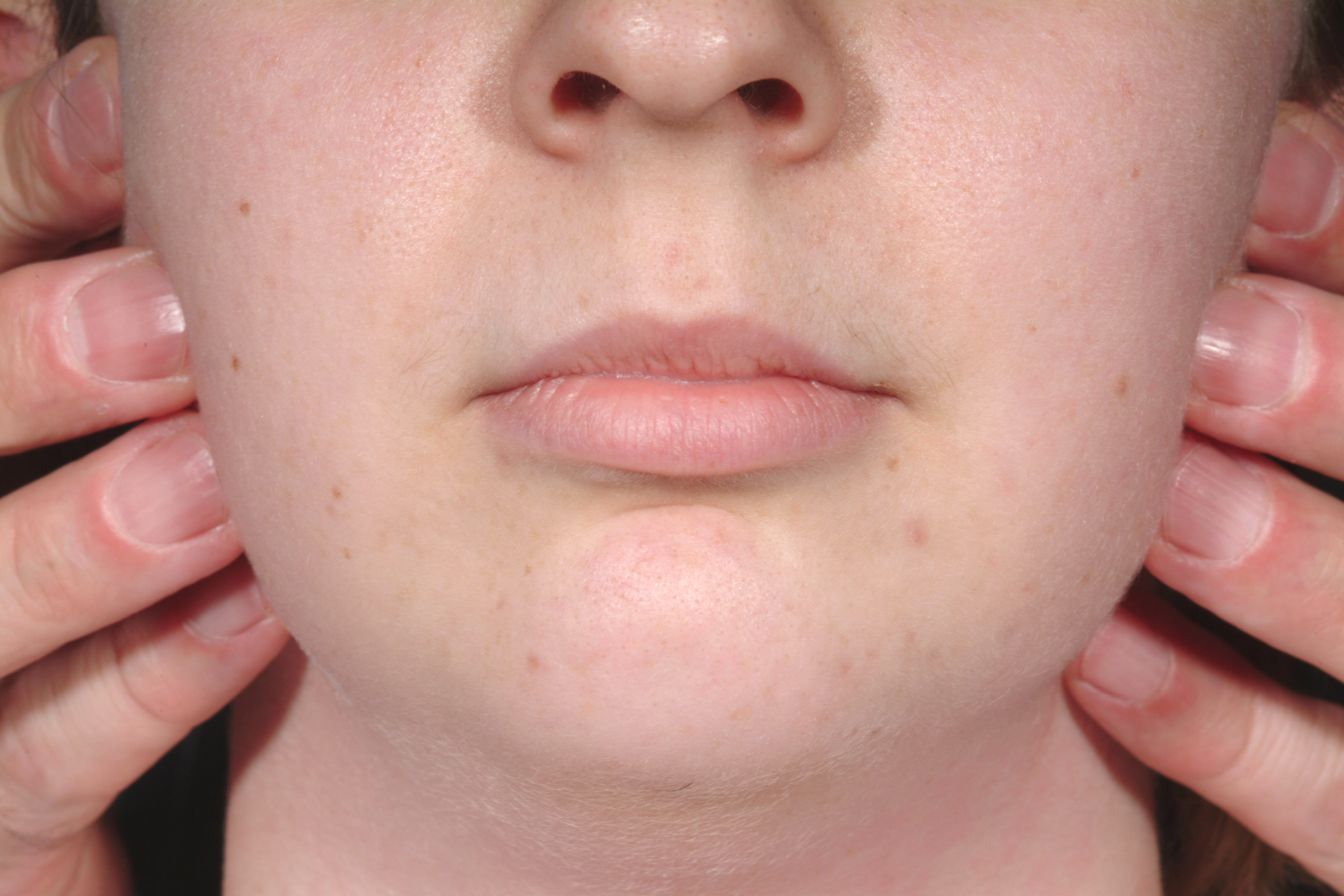
Areas of tenderness, trigger points, and patterns of pain referral should be noted. The examiner places their fingertips in the preauricular region just in front of the tragus of the ear and asks the patient to open and close the mouth – the fingertip should fall into a depression left by the translating condyle. The examination also includes listening for joint sounds. Mandibular function is assessed by noting whether the mouth opens in a straight line or deviates with jerky movements. Normal painless maximal opening should be around 40-55 mm.
Diagnosis of TMJPDS is essentially based on clinical grounds. A history of limited mouth opening, which may be intermittent or progressive, is a key feature of temporomandibular disorders.
TMJPDS needs to be distinguished from other possible conditions; differential diagnosis:
- dental pain
- disorders of the ears, nose, and sinuses
- neuralgias
- headaches
- diseases of the major salivary glands
Internal derangement
As far back as 1992 the Consensus Conference for Temporomandibular Surgery defined internal derangement as ‘a localised mechanical fault in the joint which interferes with its smooth action’. To complicate matters, however, internal derangement may also occur together with TMJPDS (see above). It is essential therefore that the contribution of each condition to the symptoms is assessed because one disorder may benefit from surgery, whereas the other will not.
Normally when the mouth opens and closes the meniscus moves in harmony with the condyle as it translates along the articular eminence. In certain circumstances, however, this gliding movement is disrupted and the disk may become adherent to the fossa either by fibrous adhesions or by the so-called ‘suction cup effect’. The disk may also change shape. As a result the mandibular condyle is unable to move smoothly and may have to ‘jump’ over the periphery of the disk in order to continue moving. This movement is felt as a click.
Why the disk becomes sticky in the first place is not known. There is, however, a mounting body of evidence suggesting that changes in the consistency of the synovial fluid as a result of inflammatory processes may be responsible. This may occur as a result of chronic pressure applied to the joint (for example, resulting from constant bruxism (teeth grinding).
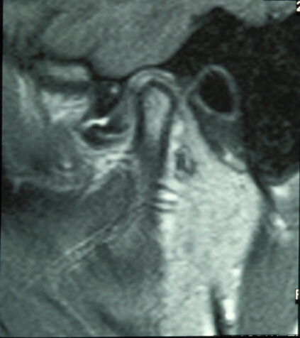
Untreated, clicking may, but not always, progress to locking, in which the disk becomes so abnormally shaped and displaced that it effectively acts like a door-stop, preventing the mandibular condyle from undertaking a full range of movements. This results in ‘locking’ of the jaw which may occur on either opening or closing of the mouth (open lock, closed lock; see Figure 2).
Pain may result from reflex muscle spasm or arise from the joint itself. It has been suggested that in susceptible patients, alterations in the synovial fluid can also result in a destructive arthropathy. These biochemical changes are similar to those seen in depression, and this helps to explain why improvement in symptoms can occur in patients who are not depressed and who take tricyclic anti-depressant drugs.
A classification system of internal derangement (based on arthrographic findings) was produced and includes:
- Class I: painless clicking; no restricted motion; slight forward displacement of the disc
- Class II: occasional painful clicking; intermittent locking; headaches; slight forward displacement of disc
- Class III: frequent pain; joint tenderness; headaches; locking; restricted motion; painful chewing; anterior disc displacement
- Class IV: chronic pain; headaches; restricted motion; early to moderate degenerative changes; flattening of eminence; deformed condylar head; sclerosis
- Class V: variable pain; joint crepitus; painful function; disc perforation; gross anatomic deformity of disc and hard tissues with degenerative arthritic changes.
Osteoarthrosis
Osteoarthrosis has been described as a degenerative (wear and tear) disease that commonly affects weight-bearing joints such as the hips and knees. However, more recently the underlying pathology has been questioned, and a degenerative cause is not so clear-cut. Osteoarthrosis is now believed to be a systemic disease with trauma only contributing on the onset of symptoms. Nevertheless, changes of osteoarthrosis can be detected in the temporomandibular joint at post mortem examination in over 40 % of middle-aged and elderly patients. Radiographically osteoarthrosis is often a common incidental finding. Despite this frequency, osteoarthrosis seldom causes symptoms. Clicking and limitation of movement are the most common symptoms; however, pain is unusual unless advanced. X-ray radiographs of the mandibular condyle show the typical changes of osteoarthrosis, namely osteophytic lipping and small subarticular radiolucent areas. As the condition progresses the articular surfaces become flattened, the articular cartilage becomes less elastic and cracks appear in the underlying bone. In localised areas there may be loss of cartilage with eburnation (a particular degenerative condition) of bone.
Symptoms include:
- crepitus
- difficulty in chewing
- reduced mouth opening
- chronic pain
Rheumatoid arthritis
This is a multisystem disease which predominately affects the joints; most commonly the hands, wrists and elbows. Progressive damage and significant symptoms tend to be uncommon in the temporomandibular joint (less than 5 %). Of these the most common symptoms are crepitus and restriction of mouth opening. Rheumatoid arthritis undergoes acute phases of exacerbation, followed by quiescent periods. Histologically there is overgrowth of the synovial lining associated with dense inflammation (pannus). This inflamed vascular mass spreads over and progressively destroys the articular cartilage, with resulting adhesions and limitation of movement. In severe cases this may progress to fibrous ankyloses (see below). In the acute phase there may be pain and tenderness over the joints, which may be hot and swollen and therefore appear infected. There may be a deep-seated, aching pain, particularly on moving the jaws. In advanced cases, limitation of movement develops. In the acute phase, an acute infection must be excluded as this is severe and potentially life-threatening; however, it is rare.
Arthritis in children (juvenile arthritis), if severe, can damage the condyle’s growth potential which can result in facial asymmetry, micrognathia (undersized jaw) and malocclusion.
Infective arthritis
With the advent of antibiotics, infective arthritis has become rare. Most cases arose from the spread of infection from the ear (otitis externa, otitis media) which is nowadays much better diagnosed and treated. However, infective arthritis is a potentially very severe condition because it may spread into the middle cranial fossa and result in intracranial sepsis.
Symptoms include:
- pyrexia and systemic upset
- throbbing pain
- swelling
- erythema
- suppuration
- very restricted mouth opening
Ankylosis
Ankylosis is an abnormal union across a joint space. This may be either fibrous or bony in origin. Commonly, ankylosis is seen in the larger joints (for instance the hips and knees following severe injuries, or the spine in ankylosing spondylitis).
Normal mouth opening is said to be greater than 40mm in adults. In addition, it should be possible to protrude the mandible at least 7mm and move it side to side a similar amount. Untreated ankylosis results in permanent limitation of the mouth opening and may restrict development of the midface if it occurs in the very young. Bony ankylosis results in the most severe limitation of opening. Ankylosis may be considered as extracapsular or (‘false’ ankylosis) or intracapsular (‘true’ ankylosis).
Causes of extracapsular ankylosis
- trauma resulting in periarticular fibrosis (e.g. lacerations, burns, dislocation)
- chronic or untreated infection next to the joint
- tumours (for example, fibrosarcoma, chondrosarcoma)
- periarticular fibrosis (irradiation, prolonged immobilisation of the jaws)
Causes of intracapsular ankylosis
- trauma (intracapsular condylar fracture, penetrating wounds, forceps delivery at birth)
- infection (otitis media, mastoiditis, osteomyelitis, pyogenic arthritis)
- systemic disease (juvenile arthritis, psoriasis, rheumatoid arthritis, osteoarthritis)
- tumours (chondroma, osteochondroma, osteoma, sarcoma, fibrosarcoma, synovial sarcoma)
- miscellaneous (synovial chondromatosis (development of surplus cartilage in the joint))
Pseudoankylosis
This condition is a mechanical interference with mouth opening which does not directly involve the joint itself. Causes include:
- trauma (e.g. depression of the zygomatic arch impinging on the coronoid or insertion of temporalis muscle)
- myositis ossificans (muscle turning into bone tissue) within any of the muscles of mastication