MRI
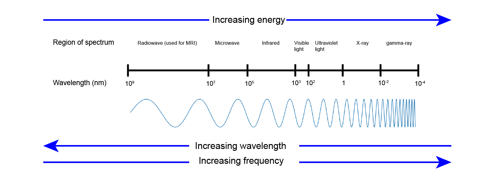
Magnetic resonance imaging (MRI) is a diagnostic scanning technique that uses a strong magnetic field (the large tube part of the scanner) and series of short bursts of radiofrequency electromagnetic waves (Figure 1) to produce three-dimensional tomographic images.
MRI looks at the behaviour of the tiny magnetic moments present in the nuclei of almost all atoms. Clinical MRI specifically looks at the behaviour of the nuclear magnetic moments of hydrogen atoms: hydrogen atoms are everywhere in our bodies, mostly in the form of water but are also present in fat, muscles, tendons, bones, nerves, etc., and the hydrogen isotope with a magnetic moment, 1H, has very favourable magnetic resonance properties. 1H is not only present everywhere in the body, it is also present in very high natural abundance (approximately 99 % of hydrogen atoms are this (stable) 1H isotope), in addition to several more general ‘MRI friendly’ properties of 1H. The latter aspects are explained in much more detail on our detailed page about MRI.
How it works in principle
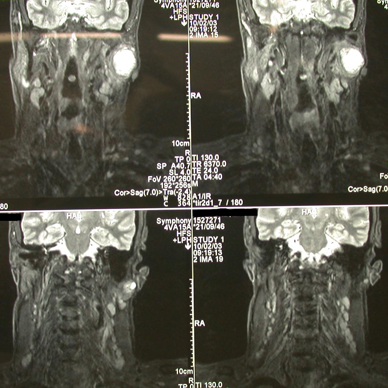
When the body, or parts of it, is placed in the strong magnetic field of an MRI scanner (inside the large tube part of the scanner), the tiny nuclei of the hydrogen atoms in 1H ‘feel’ the presence of the strong magnetic field and partially align with it (think of a strongly magnetic compass needle which aligns itself with the earth’s weak magnetic field). A short sharp burst of radiofrequency waves (a ‘pulse’) lifts this alignment, after the pulse is turned off, the magnetic moments start re-aligning with the magnetic field. While doing so, they themselves emit weak radiofrequency signals which are picked up by antennae located in the scanner. Now, that would give us a magnetic resonance signal but it would not be possible to read any information about the location of different tissues, etc, from the signal because the strong magnetic field is very uniform. Something else needs to be done to also encode information about location – a little bit like creating a giant three-dimensional jigsaw puzzle made up of thousands of small parts (voxels; think of your TV or digital camera – the two-dimensional images there are made up of thousands of small parts (pixels) too) and labelling each of the voxels with its respective location. In MRI, encoding information about location is achieved by switching on/off additional (weaker) magnetic fields, called gradients, which essentially label MRI signals with information about the location from where the signal stems. The rapid switching of these gradients are the loud banging noises during the scan. MRI scans typically follow recipes of complicated simultaneous series of radiofrequency wave bursts and switching of gradients, creating thousands of little pieces of information which afterwards are put together in a computer to construct a three-dimensional image. These three-dimensional images carry very detailed information about structures and locations, and can be examined in different views (Figure 2).
In practice, the area of the body in need of MRI scanning has to be placed in the centre of the big magnetic field. In addition, some hard plastic cover may have to be placed around the part of the body being scanned. This plastic cover carries additional radiofrequency wave equipment to enable the creation of optimum images.
The radiofrequency waves used in MRI have very low energy (Figure 1) and thus do not damage body tissues (in contrast, traditional X-rays or CT scans use high-energy (ionising) radiation). However, caution is necessary with MRI scans performed on people with certain, older metallic implants in their bodies such as pacemakers, cochlear implants, some surgical clips and some artificial heart valves. Such older metallic implants may be magnetic themselves, or may be seriously disturbed by the strong magnetic field of the scanner and/or the scanning process. Modern such devices (implanted over approximately the past decade) are non-magnetic and are thus ‘MRI safe’. Some tattoo inks containing magnetic ions (Fe ions) may have an impact on image quality.
Sometimes an injection of contrast medium, usually given in the vein in the elbow region or the back of the hand, is necessary. The purpose of an MRI contrast agent is to selectively highlight certain aspects of the scans. An MRI contrast agent typically consists of molecules with magnetic properties (different from contrast media used in X-ray or CT scans). MRI contrast agents are eliminated from the body via the kidneys, hence drinking plenty of water after the scan helps to remove the contrast medium swiftly from the body. Usually the function of the kidneys will have been assessed by a blood test ahead of an MRI scan that requires the use of contrast agent.
Typically it takes about 20 to 30 minutes to complete an MRI scan. The exact duration of a scan depends on the kind of tissue, the part of the body being investigated and the required image resolution for diagnostics. There are two main reasons why it takes quite some time to collect the data for a complete MRI scan: i) the tiny magnetic moments of atomic nuclei only yield weak signals, and ii) the response of these tiny magnetic moments in producing measurable signals is rather slow. It often takes seconds for the nuclei to return to equilibrium after a pulse and thus being ready for subsequent data production in the series of tomographic data acquired – there is a large number of data to be collected in order to have a final data set with the required spatial resolution of the information.
Magnetic resonance research laboratories all over the world search for methods to boost and/or accelerate the response of the tiny nuclear magnetic moments so that hopefully in the future MRI scans will take considerably less time to complete.
An MRI scan is a painless procedure. But it is necessary to be able to keep still and lie quite flat during the scan so that clear images can be recorded (infants may have to be sedated for the recording of an MRI scan). Some people find the loud banging noises of the scanner unpleasant but earplugs and headphones are on offer to minimise the noise disturbance. Some people find confinement in the rather narrow scanner, together with additional equipment placed close to the head, a little claustrophobic. The best advice really is to relax and ignore this environment as best one can – it is not in any way dangerous, just not particularly ‘nice’. Recently MRI scanners with open magnets have become available. These scanners may feel less claustrophobic but they deliver lower quality images.