Broken bone
Fracture
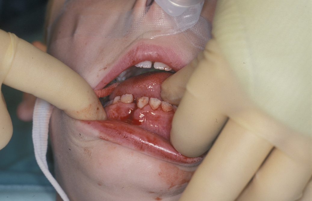
In general a facial fracture is recognisable clinically based on symptoms of pain, malocclusion (misalignment between teeth in upper and lower jaws) and diplopia (double vision). There will be signs of malocclusion, visible deformity, sublingual (underneath the tongue) haematoma, subconjunctival haemorrhage (‘complete’ red eye), epistaxis (bleeding from the nose) and abnormal mobility. Figure 1 shows a typical case of a mandibular (lower jaw) fracture.
Confirmation of site and specific fracture pattern details are obtained from X-ray images (by plain films at right angles) and CT scans . The imaging will help the surgeon plan the operation, find, realign the fragments and avoid missing a fracture segment when operating. 3D reformatted images from CT scans can be helpful in the course of treatment: it is increasingly available and may be helpful in demonstrating injuries to patients and support staff. Additional suspected or evident injuries to soft tissues such as ligaments need to be investigated by MRI scans.
Locations of fractures; incidence and aetiology
Mandibular fractures (fractures of the lower jaw)
Fractures of the mandible account for 20% of all facial bone fractures. 80% of these are in males.
In Britain 75% of mandibular fractures are due to interpersonal violence, usually involving alcohol. Sports injuries (10%), road traffic collisions (8%) and falls (4%) are the next most common factors. This varies internationally, particularly in relation to the availability of intoxicants and the presence or absence of civil unrest. A study in Aachen, Germany on mandibular fractures with cases between 1995-2007 and n = 370 patients revealed 40% interpersonal violence, 20% falls, 14% road traffic collisions, 10% bicycle, 5% sports injuries, suggesting variation even within Northern Europe.
Zygomatic complex fractures (fractures of the cheek bone)
A zygomatic complex fracture is a fracture of the zygomatic bone (zygoma). The zygomatic bone forms the cheek bone prominence, the lateral orbital wall and the lateral aspect of the orbital floor (socket of the eye), and the buttress of the maxilla (upper jaw). Because of this, fractures in this region are often referred to as the zygomatico-orbital (and sometimes zygomatico-maxillary) complex fracture. The zygoma also has a posterior extension (to the back; temporal process) which joins the zygomatic process of the temporal bone to form the zygomatic arch. Fractures in this region are referred to as zygomatic arch fractures.
Fractures of the zygoma account for 25% of facial fractures and are the second most common. 80% occur in males.
35% of the fractures result from interpersonal violence, 25% from road traffic accidents, 20% from sports injuries, and around 10% from falls. Again this distribution varies internationally.
Nasal fractures
Fractures of the nasal bones account for 50% of all facial fractures. They are often regarded as trivial injuries and poor outcomes of some of the more displaced fractures are seen as a result of this.
40% of nasal fractures are caused by interpersonal violence. Falls (25%), sports injuries (20%) and road traffic collisions (10%) account for the rest.
Midface fractures
Midface fractures are less common than the other bony injuries, accounting for 5% of facial fractures in the UK. The introduction of seatbelt and crash helmet legislation and improved industrial health and safety regulations have been held responsible for this. In countries where this legislation is not enforced (such as the Middle East and India) a higher incidence is seen.
Road traffic collisions are still the cause of around 50% of all mid-face injuries; with falls (20%), sports injuries (15%) and interpersonal violence (10%) accounting for the remainder. The force required to create these injuries accounts for the differences seen between midface fractures and the more common injuries.
Frontal bone fractures and frontal sinus (space behind eye brow region, connecting bones of skull and face) fractures
These are relatively rare fractures and are seen by both neurosurgery and maxillofacial surgery. These fractures will often be part of a more complex set of damage from serious head injury. Data regarding incidence is sparse and inaccurate.
These fractures involve considerable force, usually resulting from road traffic collisions, industrial accidents and assaults.
Craniofacial fractures (combined fractures of facial and skull bones)
These fractures are complex in nature and require treatment by a multidisciplinary team. Craniofacial injuries may be associated with a significant head injury and further detailed imaging investigations for assessment are important. It is imperative that the brain is covered to prevent further injury.