Oral mucosal lesion
Contents
Oral mucosal lesions related to infections
Bacterial, fungal and viral infections are common causes of oral mucosal lesions. In fact, infections of the mouth are so common that they are described and discussed in a separate section dealing with infections. Here we list common infections which typically give rise to mucosal lesions.
Oral bacterial infections include pericoronitis, alveolar osteitis (dry socket), acute and (more commonly) chronic periodontal disease, necrotising ulcerative gingivitis, dentoalveolar abscess. Scarlet fever, syphilis and tuberculosis can all give rise to oral lesions.
Viral infections with oral manifestations include herpes simplex (acute, chronic and recurrent), varicella zoster (primary causing chickenpox, later in life after dormancy causing shingles), Epstein-Barr virus (glandular fever), cytomegalovirus, coxsackie virus A (herpangina and hand, foot and mouth disease (affecting children)) and human immunodeficiency virus, HIV infections (increased risk of oral bacterial and fungal infections).
Fungal infections are almost always caused by candida species (albicans being the most common), causing acute (thrush) and chronic forms of fungal infection (candidiasis) and include opportunistic infections following treatment with broad-spectrum antibacterials or inhaled steroids (erythematous candidiasis). Chronic hyperplastic candidiasis can give rise to a form of candidal leukoplakia, persistent white patches with a tendency for abnormal cell growth (see below).
Recurrent aphthous stomatitis (mouth ulcers)
Minor aphthous ulcers are a very common condition, affecting about a third of the population, and are the most common form of mouth ulcer. The most likely cause is a combination of some (auto)immune reaction, possibly in combination with some genetic disposition; the condition occurs in familial clusters.
The small ulcers (a few mm in size) tend to appear in groups in the oral mucosa and heal, without scarring, in 1 to 2 weeks. Recurrence intervals are usually a few months, the condition is worsened by stress and is influenced by hormonal effects (such as the menstrual cycle). Occasionally such minor ulcers can be signs of other diseases (see below).
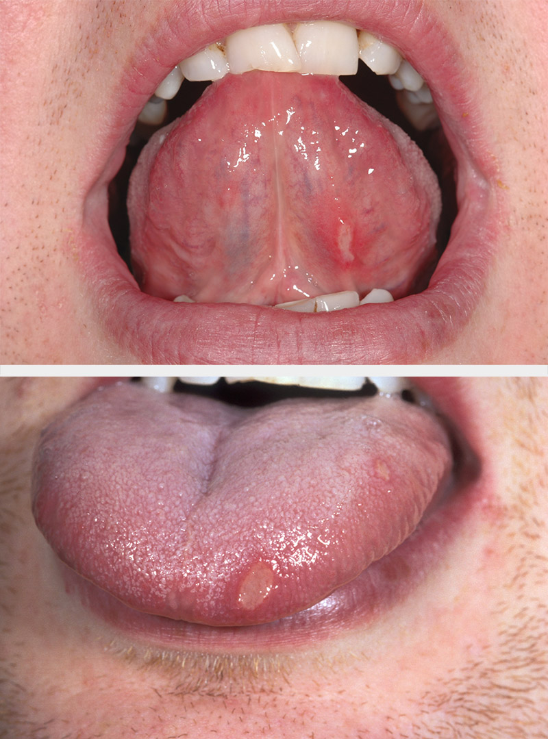
Major aphthous ulcers are a less common variant of mouth ulcers (about 10 % of cases), with more severe symptoms, larger ulcers and more frequent recurrences. These ulcers heal with scarring and are more often a sign of some other underlying disease (see below).

The least common form, herpetiform ulcers, mostly affect older females. Herpetiform ulcers resemble oral ulcers caused by viral liver infections (see below) but are not caused by infection. These ulcers occur in clusters of numerous small, painful ulcers in any area of the mouth and frequently recur over extended periods of time (sometimes several years).
Mucositis
Oral mucositis is a severe and painful inflammation and ulceration of the mucosa, the lining of the mouth. Mucositis can affect the entire gastrointestinal tract and is a common severe side effect of cancer treatments, both radiotherapy and chemotherapy, as well as part of a rare but serious complication after bone marrow transplants (so called graft versus host reaction). Oral mucositis can be so severe as to be a treatment-limiting effect. The mucosa is vulnerable to these effects because of its relatively high rate of tissue renewal.
Mucositis ulcers are vulnerable to infections and necrosis. Some degrees of oral mucositis occur commonly as an unwanted effect of nearly all systemic chemotherapies (with some agents, such as 5-fluorouracil, more likely to cause severe mucositis than other cytotoxic drugs) and radiotherapies. It often is particularly severe during leukaemia treatment in children (childhood leukaemias tend to be treated with aggressive chemotherapy schemes).
Mucositis caused by radiotherapy for the treatment of head and neck cancers, especially in combination with chemotherapy, tends to be particularly severe. Radiotherapy applied to the head and neck region almost always causes (variable degrees of) damage to the salivary glands, leading to problems with dry mouth, xerostomia which exacerbates the mucositis.
Oral mucositis is classified in four degrees of severity, with class 3 (only liquids can be swallowed) and class 4 (nothing can be swallowed) calling for immediate intervention by alternative feeding regimes, if not hospitalisation. There is an ongoing debate about these approaches, about the optimal overall duration, various kinds and different starting points of such feeding interventions.
Vesiculo-bullous lesions (blisters)
These are technically slightly different lesions from ulcers. Ulcers are lesions of the mucous membrane, small (vesicles) and large (bullae) blisters are lesions of the epithelium (made up by superficial cells of the thin surface lining tissue). Blisters can either interrupt the adhesion between epithelial cells (intraepithelial blisters) or detach the epithelial layer from its underlying tissue (subepithelial blisters). These intraepithelial and subepithelial blisters / lesions are by and large oral manifestations of a number of different underlying diseases (see below), with the important exception of the spontaneous formation of oral blood blisters of unknown cause (angina bullosa haemorrhagica, these are irritating but benign in nature).
Persistent patches and lesions
A number of persistent white (leukoplakia), red (erythroplakia) and speckled (erythroleukoplakia) patches, submucous fibrosis (scar tissue formation beneath the mucosa) and a rare variant of lichen planus (see below) all belong to a group of oral lesions that are associated with variable degrees of abnormal cell growth (dysplasia) and a possibility for malignant transformation of the lesion. These lesions are therefore sometimes called premalignant lesions; we discuss dysplasia in a separate section:
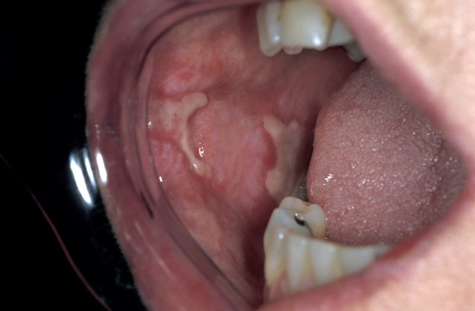
Lichen planus is a common skin condition (estimated 1 % of the world population), thought to be an immune disorder. It is an itchy non-infectious rash mostly affecting people older than 40 years, women are more often affected than men. Approximately in half of the cases not only the skin but also the oral mucosa is affected. Lichen planus may exclusively affect the oral mucosa.

Oral lichen planus typically exhibits white or reddish lace-like patterns on the surface of the tongue or inner cheek; if ulcers develop these may be painful. A biopsy may be necessary to confirm the diagnosis. A rare (approximately 1 %) sub-variant of oral lichen planus, erosive lichen planus (see Figure 4), is thought to belong to the group of potentially premalignant lesions whereas for the more common forms no such association is known. Oral lichen planus may persist for many months or years.
Frictional keratosis is characterised by white patches caused by some minor local trauma, such as ill-fitting dentures or a sharp tooth.
Smoker’s keratosis are white patches caused by irritation by chemicals in tobacco smoke.
Oral cancer
The majority of oral mucosal lesions are benign, but it is definitely a good idea to carefully check any persistent lesions in order to exclude malignancy. That may require taking a biopsy.
Oral mucosal lesions related to other diseases
Many non-oral diseases have oral manifestations; these can be early or late signs and symptoms of other diseases. If oral manifestations happen to be an early sign of some other disease, it can (and does) happen that this disease gets initially, or tentatively, diagnosed in a maxillofacial clinic.
Apart from a wide range of infections (see above), in particular many skin diseases, haematological (blood) disorders and disorders of the gastrointestinal system lead to oral manifestations.
Skin diseases with oral manifestations include
- lichen planus (see above);
- dyskeratosis congenita (rare inherited condition, leading to bone marrow failure; oral leukoplakia and periodontal disease common);
- dermatomyositis (inflammatory condition affecting muscles and skin; may cause mouth ulcers or inflammation of gums);
- systemic lupus erythematosus (systemic autoimmune disorder, may give rise to mouth ulcers or rashes);
- pemphigus (rare chronic skin disorder, probably an autoimmune condition, that causes blisters and sores of skin and oral (and genital) mucosa);
- dermatitis herpetiformis (chronic inflammatory condition that affects skin and mucosa; leads to blisters);
- epidermolysis bullosa (group of skin and connective tissue disorders, large blisters as oral manifestation, may cause swallowing difficulties, caries a common problem);
- erythema multiforme (immunological skin reaction to mostly infections or medications, tends to include oral blisters);
- Ehlers-Danlos syndrome (rare inherited connective tissue disorder, periodontal disease and bleeding of oral mucosa common);
- Darier’s disease (rare inherited skin condition, papules and white patches on gums and palate).
Haematological disorders with oral manifestation include
- anaemia (caused by nutritional deficiencies (mainly iron, vitamin B12 and folic acid), recurrent oral ulcers);
- haematological malignancies (different forms of leukaemia, lymphoma and myeloma) typically have a range of oral manifestations. This is discussed in our section about haematological malignancies.
Gastrointestinal disorders with oral manifestations include
- coeliac disease (commonly known as gluten intolerance; may cause recurrent oral ulcers alongside sore tongue and lips);
- ulcerative colitis (long-term inflammation of intestines with mostly gastrointestinal signs and symptoms, but may include oral lesions and lumps);
- Crohn’s disease (inflammatory bowel disease of unknown cause; may affect all parts of gastrointestinal tract, including mouth ulcers).
Other conditions with oral manifestations include
- Behçet’s disease (rare systemic inflammatory condition of the blood vessels; amongst many other signs and symptoms, most common painful mouth ulcers);
- renal failure (may cause white oral patches);
- allergies (offenders include dental materials, a wide variety of foods and a wide range of drugs (see below)).
Oral mucosal lesions related to drugs
The list of medicinal and recreational drugs causing, for example, difficulties with dry mouth, xerostomia is long and essentially includes every type and class of drug. Similarly, the list of drugs giving rise to other oral lesions is also long and varied, below some examples:
- pigmented patches – often caused by recreational drugs, including coffee/tea, paan, nicotine, especially in conjunction with suboptimal oral hygiene, chlorhexidine (mouthwash);
- chemical burns – what it says on the tin, harm caused by direct contact of mucosa with strongly acidic or other chemically reactive substances; misguided use of aspirin tablets (pressed against aching tooth), trichloroacetic acid (haemostatic and etching agent used in dental practice), sodium hypochlorite (bleaching agent, widely used in root canal treatment) and phenol (component in some mouth rinses) can cause such lesions;
- serious oral ulcerations and/or agranulocytosis (agranulocytosis is an acute and serious lack of particular white blood cells, with high risk of serious infections and causes severe oral ulcerations) – drugs suppressing bone marrow function, phenytoin (anti-epileptic drug), chloramphenicol (antibacterial agent), cisplatin, 5-fluorouracil, busulfan and other chemotherapy drugs, captopril (anti-hypertension drug) can cause agranulocytosis and/or other forms of severe oral ulcerations;
- exfoliative stomatitis (oral manifestation of skin-shedding as a drug reaction) – this potentially serious drug reaction has been associated with gold drugs (arthritis treatment), antibacterials (sulphonamides, penicillin and its derivatives, isoniazid), blood pressure medications (Ca channel blockers), anti-epileptic drugs (phenytoin and others), barbiturates (sedation) and a number of topical medications;
- gingival hyperplasia (enlargement of the gums) – Ca channel blockers (anti-hypertensive, blood-pressure lowering drugs) and phenytoin (anti-epileptic drug) common offenders;
- allergies with oral manifestations – common offenders amongst systemically administered drugs are some antibacterial agents, β-lactams (penicillin and its derivatives) and some analgesics (NSAIDS); locally common offenders are components of toothpastes, in particular benzoates (preservatives), sodium lauryl sulphate (foaming agent) and artificial flavourings.