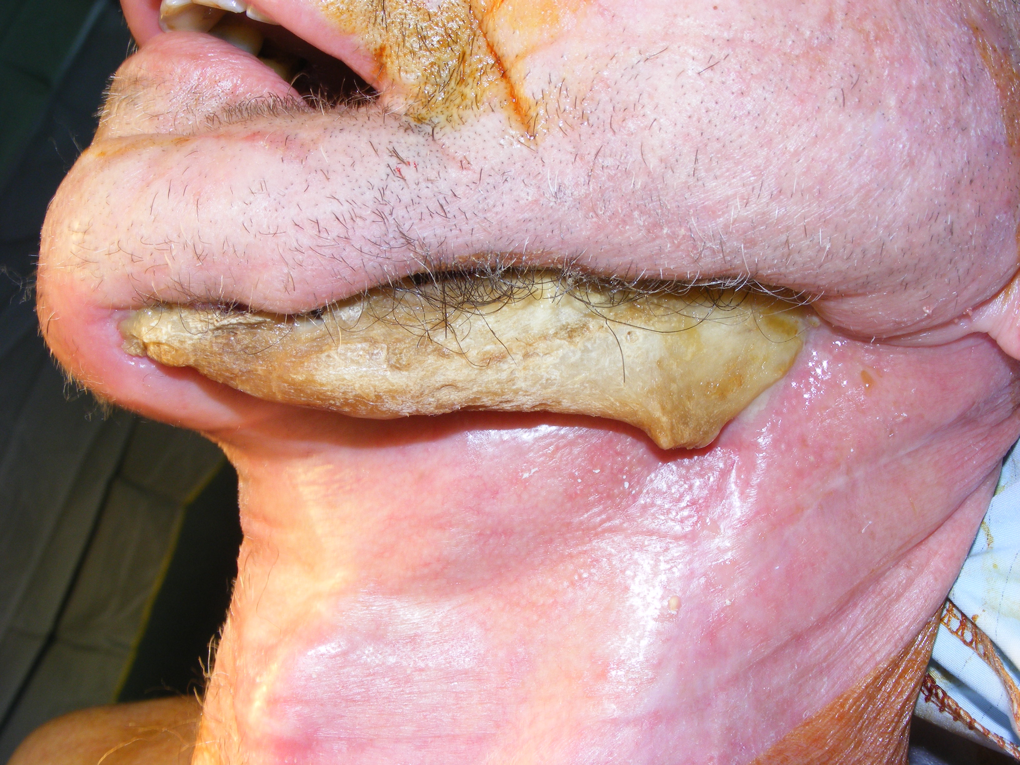
Mouth cancer
Cancer of the oral cavity includes cancer of the lip, tongue, gingivae, floor of the mouth and palate. Of these, the commonest sites are of the tongue and floor of the mouth.
Squamous cell carcinoma forms some 90% of oral cancer. Other types of oral cancer include those arising from salivary glands, bone and other connective tissues, melanoma and lymphoma.
There are about 6,200 new cases each year in the UK. The incidence of oral cancer is increasing. Globally, cancer of the mouth and pharynx is the 6th commonest type of cancer. Although less common in the United Kingdom (head and neck cancer is 12th most common cancer for males), it is the commonest in the Indian sub-continent (India, Pakistan and Bangladesh). There is a north-south divide in the UK, with higher incidence recorded in the northern parts of England, Scotland and Northern Ireland compared to the more affluent south.
Oral cancer is more common in males. The vast majority of patients with oral cancer are middle aged or older (98% over 40 years old). It is, however, increasing in younger patients, in particular, non-smokers. Possible reasons given for this rise are the increased consumption of alcohol, and involvement of the high risk strains of the Human Papilloma Virus (HPV 16 and 18). This is an established cause of a rapid increase in oropharyngeal cancer (which is considered a separate cancer from oral cancer in some epidemiological data), particularly that of the tonsil and base of tongue which are more frequently treated primarily non surgically although this has led to a different series of post treatment problems.
Head and Neck Cancer Staging
This is a process whereby the extent of the cancer is assessed and placed into a category. Staging is used to compare treatment and prognosis results across different treatment centres and tactics. It involves combining several diagnostic tests, clinical examination, imaging techniques MRI, and/or CT and sometimes PET/CT. In reality although a cancer is always given a stage using the Tumour, Node, Metastases (TNM) system (UICC staging) the degree of inter and intra centre variability is high and treatment intent and approach is always personalised to some extent.
Diagnostic tests
These include blood tests such as full blood count, urea and electrolytes including an EGFR (kidney function) to ensure contrast enhanced scans can take place, grouping of blood type, liver function tests and others. MRI of the head and neck area, CT of the chest and abdomen and PET/CT are regularly carried out. Plain radiographs (X-ray) of the jaws even if no teeth are present are taken (there may be buried tooth roots not obvious and if the jaws are exposed to radiotherapy treatment surgery afterwards creates a risk of a serious condition called osteoradionecrosis.
The principle issue here is that any teeth which need to be removed before radiotherapy should be removed either at the time of surgery or 3 weeks before radiotherapy starts. A restorative dentist is usually involved.