Surgical endodontics
Contents
General comments on indications and contraindications for surgical endodontics, success rates and alternative treatment by dental implants are given. Some specific procedures (including incision and drainage, perforation repair, root and tooth resections, extraction with replantation, revision surgery) and aspects depending on specific areas of the mouth are outlined.
Indications and contraindications for surgical endodontics
The consensus report of the European Society of Endodontology suggests that endodontic surgery is indicated when:
- a canal is obstructed, for example with a separated instrument, and there are associated symptoms;
- extruded material is present with clinical symptoms, such as pain, that continue over a significant time period (at least one week);
- conventional root canal treatment has failed and retreatment is inappropriate;
- perforation of the root or the floor of the pulp chamber has occurred, with radiological findings or clinical symptoms, and where repair from within the pulp cavity is not possible.
In almost all situations, conventional root canal treatment should be attempted first, and after a sufficient period of time the tooth is reassessed for further treatment. If the non-surgical root canal treatment has been performed to the highest practicable standard but symptoms persist, then surgical endodontic therapy may be appropriate. If, however, there have been procedural errors or there are technical deficiencies in the conventional root canal therapy, then conventional retreatment is indicated prior to considering surgery.
In some situations, it is impossible to revise the existing root filling, particularly when some of the older pastes have been used, even though ideally one would wish to do so. In these circumstances endodontic surgery is the only viable option to avoid extraction if symptomatic, but success rates are low.
Where a tooth has been restored with a post crown, the principles outlined above still apply, and attempts should usually be made at dismantling the post crown and instigating conventional retreatment. Occasionally, however, removal of a post may carry a significant risk of root fracture, and in such cases, it may be preferable to leave the post in situ and carry out surgical endodontic therapy.
Contraindications to surgical endodontic treatment include:
- anatomical factors such as an inaccessible root-end;
- a tooth with a hopeless prognosis (for example, advanced loss of periodontal support) that cannot be made functional or does not contribute to aesthetics;
- a patient who does not provide informed consent to undergo the procedure;
- a patient with a compromised medical history, such as uncontrolled systemic disease (for example, diabetes or some haematological disorders).
Success rates
For non-surgical endodontic treatment, success rates of 86 to 96 % have been reported, and for retreatment 60 to 98 %. Clinical success following surgical endodontic therapy has been reported to vary between 58 and 96 %. The wide range of reported success rates may be due to differences in evaluation criteria, diagnoses, operator skill and duration of follow-up periods.
Bacteria are the main cause of periradicular disease, with enterococcus faecalis having been found to be the predominant microbial in failed cases and actinomyces species being able to survive in periapical tissues. An effective coronal seal is thought to improve success rates by reducing the viability of bacteria in the root canal system and the apical area.
Factors thought to improve success include:
- position of the tooth in the arch, favouring anterior teeth;
- a well condensed orthograde root canal filling;
- a small apical lesion (less than 6 mm);
- use of an ultrasonic microtip;
- first attempt at apical surgery for the tooth;
- use of an endoscope.
Alternative treatments
There are a number of ways in which non-vital teeth may be preserved. However, there is an increasing body of evidence supporting the long term viability of dental implants. Accordingly, atraumatic removal of such teeth with preservation of the maximum amount of healthy bone and immediate implant replacement may become the standard treatment for non-vital teeth which cannot be salvaged by conventional or surgical endodontics. It should be remembered that endodontic treatments, conventional and surgical, are perfectly valid techniques to preserve natural teeth and cost considerable less than dental implants.
Specific procedures
Incision and drainage
Incision and drainage are one of the few surgical procedures which should be performed in the presence of acute inflammation. The main indication for incision and drainage is the presence of pus within the tissues that cannot be drained through the root canal. Anaesthesia is obtained with the use of surface analgesics, which can be supplemented with a few drops of local anaesthetic solution injected submucosally. An incision is made with a number 11 scalpel blade (a blade with a sharp pointed tip), a microbial swab is taken and drainage affected. Alternatively, a wide bore needle and sterile syringe can be used to aspirate the contents of the swelling and culture and sensitivity testing carried out. In practical terms it will be days before the results of these microbiological tests are available. If there is no drainage, antibacterial agents such as a penicillin and/or metronidazole should be used. Significant infection beyond the confines of the dentoalveolar complex mandates aggressive antibacterial treatment in addition to adequate drainage.
Treatment of specific areas of the mouth
Specific areas of the mouth pose various difficulties during apical surgery, and an understanding of the local anatomy is of immense importance when considering surgery.
In the treatment of upper anterior teeth, reflection of the flap to provide adequate access to the apex of upper central incisor teeth can be compromised by a prominent and/or low anterior nasal spine. Extension of the flap around more teeth and increasing the size of any relieving incisions often allows sufficient access to perform the surgery. The apices of upper lateral incisors can be deeply placed in the alveolar bone, which makes identification of the apex and access for apical surgery more difficult. The roots of upper canine teeth are occasionally very long and access to the apex may be restricted by the buccal reflection. Again, adequate relieving incisions help overcome this.
The maxillary sinus is often in close relation to the apices of upper posterior teeth, but a careful surgical technique will avoid damage to the sinus. It is advisable to burr down the root apex to the desired level, rather than resecting it, so that the resected tip cannot be displaced into the sinus or under its lining.
Apical surgery on upper first premolar teeth can be challenging because of the deeply placed palatal root. It may be necessary to resect a greater length of the buccal root than normal to provide sufficient access to the palatal root. The upper second premolar tooth is usually single-rooted, but there may be two separate root canals or two canals joined by an isthmus. In these situations, it is important that both canals, together with any isthmus of tissue that may be present, are sealed. Access to the palatal root of upper molars is almost impossible, but fortunately the palatal root is readily treated by conventional endodontics. The buccal roots are normally fairly superficial but occasionally they are deeply placed beneath the zygomatic buttress.
The apices of the lower anterior teeth are usually deeply placed in the mandible, which makes access extremely difficult. In addition, the mentalis and buccinator muscles are attached at a relatively high level, so that reflection of the flap to the level of the apices can be problematic. Therefore, a rectangular flap design with two relieving incisions is advised, and a small bevel of the root face may be necessary so that the canal(s) can be visualised. Local anaesthesia can also be surprisingly difficult, possibly due to sensory innervation by the nerve to mylohyoid.
The mental nerve usually emerges from the mental foramen below and between the lower premolar teeth. Therefore, vertical relieving incisions should be avoided in this area. Also, during root resection, it may be desirable to resect the root at the desired level, rather than burring it down, so as to reduce the risk of damage to the mental nerve. Whether or not apical surgery is possible on lower molars depends upon the degree to which the corner of the mouth can be retracted. If adequate retraction can be obtained and the attachment of the buccinator muscle is sufficiently low, surgery may be possible. A wide flap is usually required to enhance access, and it is often necessary to remove a considerable thickness of bone to gain access to the roots. Care is needed to avoid damage to the inferior alveolar nerve, and again it is advisable to resect the root rather than burring it down.
Corrective endodontic surgery
Corrective surgical endodontics includes:
- perforation repair;
- root resection;
- tooth resection;
- extraction with replantation.
Many of the basic surgical principles for ‘apical surgery’ can be applied here, for example regarding flap design, haemostasis, and root-end filling materials.
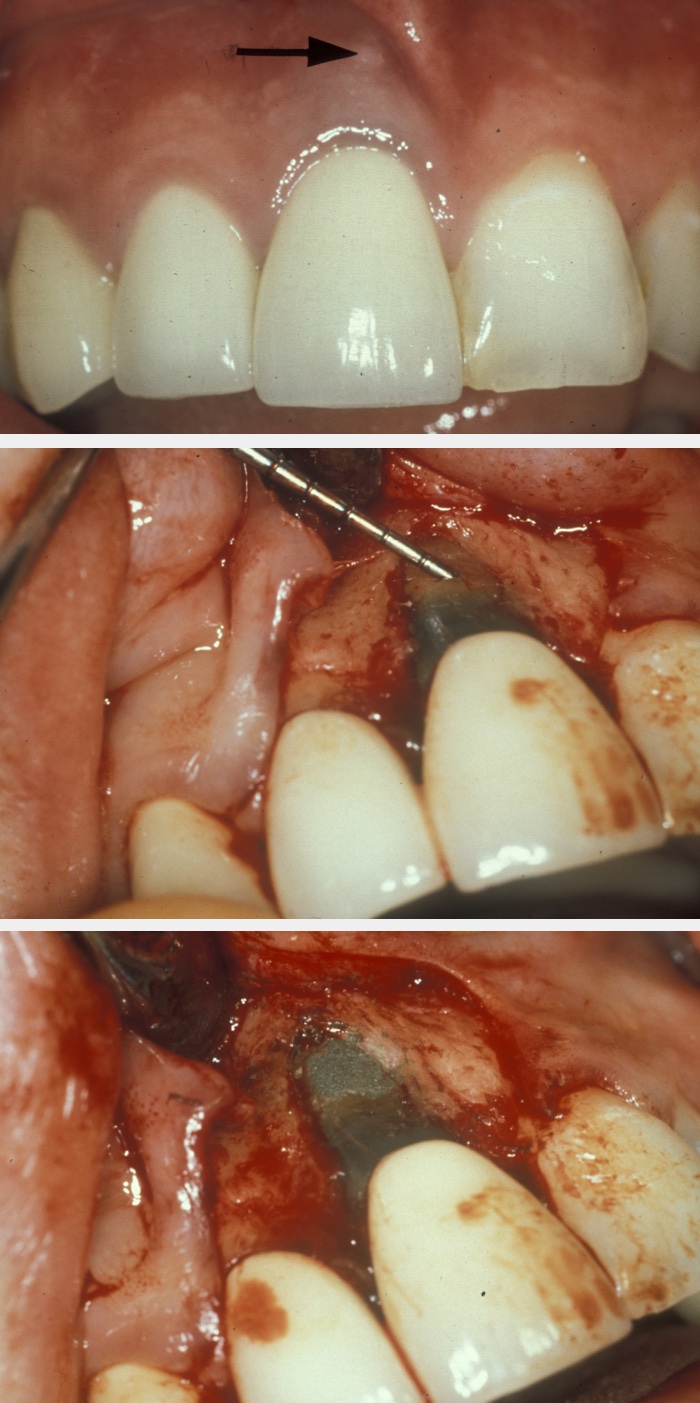
Perforation repair becomes necessary to deal with root perforations caused by broken endodontic instruments or damage by posts, and resorption cavities. If the prognosis of the tooth is acceptable, surgical access is gained to the site of the perforation, the area is prepared and then filled with, for example, mineral trioxide aggregate. Figure 1 illustrates an example; the apparent over-bevelling is a consequence of this being a perforation repair and not a conventional apicectomy.
The objective of root resection is to remove an entire root or roots from a multi-rooted tooth, but without removal of any portion of the crown. The main indications are when one root is affected by advanced periodontal disease but the rest of the periodontium is relatively unaffected, or where it is impossible to carry out root canal therapy in a root.
A suitable mucoperiosteal flap is reflected and the root is exposed by removing the overlying bone. The root is then resected from the tooth, removed, and the portion of the crown adjacent to the resected root re-contoured to make the site easily cleanable. The flap is sutured.
In tooth resection, the coronal tooth structure as well as the root is cut. The procedure is mainly performed on mandibular molar teeth with furcation involvement. The separated part of the tooth may either be removed or left in situ and restored. In the latter case, a molar is effectively converted into two premolar units.
Extraction with replantation involves intentionally removing the tooth from its socket, carrying out the surgical endodontic procedure of choice (for example apical surgery, perforation repair) extra-orally, and replanting the tooth. Although not commonly performed, extraction with replantation is useful when access for surgery in situ is impossible and the alternative would be loss of the tooth. There is a risk of damage to the tooth during the extraction process, which may preclude the procedure. This technique has largely been replaced by immediate use of dental implants.
Revision surgery
The cause of failure of endodontic surgery should be identified before considering revision surgical endodontics, and an assessment should be made of the likelihood of correcting this. Success rates for repeat apical surgery are in the region of 30 to 40 %, which is much lower than for the initial procedure.
Root canals that have not been adequately sealed are the commonest cause of failure of both conventional and surgical endodontic therapy. If surgical treatment has been performed in the absence of adequate cleansing and obturation of the root canal system, failure is to be expected. Repeat surgery will only be successful in these situations if the principles of conventional endodontic treatment are carried out first to ensure the best possible coronal seal. Failure caused by an inadequate apical seal can be rectified by ensuring there is no contamination of the root end cavity at the time of surgery, and by using an appropriate root end filling material.
Sometimes, during apical surgery root canals are incompletely sealed surgically because an additional root has not been identified. This should be suspected if, at the time of repeat surgery, the apical seal in the treated root appears intact and the surrounding tissues have healed. Radiographs taken at different angles can be useful in identifying additional roots. Often it is the deeply placed palatal root that is missed, particularly of upper premolar teeth. An incomplete seal may also be present when there are two portals of exit of the root canal in the same root and only one has been treated. Alternatively, if both canals within a single root have been sealed but the intervening isthmus of tissue has remained untreated, failure can ensue.
From a practical view point, revision surgery is more difficult, particularly when raising a flap that is scarred from previous surgery. The cause of the failure will determine which procedure is undertaken, but often the retrograde seal requires replacement. Care is needed when removing the old material to ensure it does not scatter unnecessarily in the surgical field. Depending on which material is used, these small fragments can cause staining of the mucosa or a foreign body reaction in the tissues. Consideration also needs to be given on whether the root is resected further at the time of second surgery. In most cases it is acceptable to reduce the root length a further 2 to 3 mm during revision surgery. However, in the presence of periodontal pocketing, resection of too great a length of root will reduce the distance between the epithelial attachment and the root end. Therefore, a small amount of further periodontal breakdown and migration of the epithelial attachment will result in a full-length pocket from the gingival crevice to the apical tissues.