Soft tissue necrosis
Débridement
Débridement is the technical term for removal of necrotic (and/or infected) tissue in order to expose viable healthy tissue to enable healing of a wound and to prevent development and spreading of infection(s) of a wound. There are a variety of ways to achieve removal of necrotic tissue, depending on the cause and extent of the necrosis.
It is necessary to remove necrotic tissue from a wound because its presence maintains the initial inflammatory response of the body to damage, preventing progression to the wound-healing phase. Necrotic tissue is also a good breeding ground especially for bacterial infections. The blood supply to necrotic tissue is usually impaired, so that systemic or topical antibacterial medications may not reach their targets and remain ineffective. Eradication of infection in such wounds thus almost always necessitates surgical débridement (see below).
Débridement can be achieved in different ways. For small and non-infected superficial necrotic areas of skin it may be possible to exploit the body’s own enzymes to dissolve any debris and dead tissue. This is referred to as autolytic débridement and usually needs to be supported by special wound dressings, films and gels that keep the necrotic tissue in contact with body / wound fluid. If this is an option, it is a highly selective and gentle mechanism as only the necrotic tissue will be liquefied in this way.
Débridement of superficial necrotic skin wounds by relying on the digestion of this dead tissue by other enzymes is an alternative. A range of such enzymes, isolated from plants (such as bromelain) or microorganisms (such as clostridium histolyticum) exists. They are typically less selective than the body’s own enzymes and are of varied effectiveness. This approach is not a standard treatment.
There are some further non-standard treatment options for superficial necrotic skin wounds. These include application of manuka honey; this could be considered a special case of application of digestive enzymes (see Figure 1). Another highly selective form of treatment is the use of maggots. These creatures feed exclusively on necrotic tissue and thus provide a selective and pain-free form of wound débridement (the use of maggots for such purposes is probably better known for the treatment of non-healing diabetic foot ulcers).
Surgical débridement
Surgical débridement is the fastest and ‘cleanest’ way to remove necrotic tissue, to strip a wound back to viable tissue, and to enable and improve healing. Viable tissue here means tissue with a healthy blood supply. Surgical débridement may be carried out using traditional surgical instruments such as scalpel or scissors, or a laser. It is a selective method with a high level of control of the procedure, allowing in-depth inspection of necrotic and adjacent tissues. The area surrounding the necrotic tissue is thoroughly cleaned and disinfected, the wound is carefully probed and inspected before all necrotic tissue is removed. It may be necessary to repeat the procedure and the presence of bacterial infection(s) will necessitate the use of systemic antibacterial medications in addition to surgical intervention. The choice of the most suitable antibacterial drug(s) depends on the infecting microorganism(s), where infections with MRSA, a range of Gram-negative and/or anaerobic bacterial strands, or some streptococci strands are the prime considerations.
Altogether the surgical management of soft tissue necroses is very similar to the surgical treatment protocols for serious abscesses and other soft tissue infections.
Hyperbaric oxygen therapy, HBO may be another option to support surgical and antibacterial treatments for some soft tissue infections and necrosis. However, the evidence base is conflicting. While there is some (weak) evidence of occasional beneficial effects, there is a complete lack of criteria that would allow one to predict with confidence in which circumstances HBO may be beneficial.
Surgical resection and reconstruction
In a maxillofacial context, a number of causes can lead to severe and persistent soft tissue necroses in need of more radical surgical interventions to achieve healing and preservation of function. Examples are soft tissue necroses developing after radiotherapy treatment, or severe infections involving deeper tissues such as necrotising fasciitis.
Radical resections in the maxillofacial region for the eradication of soft tissue necroses have similar restrictions and reconstructive requirements as do other radical resections. For example, tissue necrosis and destruction may be near to vital structures, a particular concern in the head & neck region.
Some considerations for the surgical resection of soft tissue necroses in the maxillofacial region are specific to the respective underlying cause.
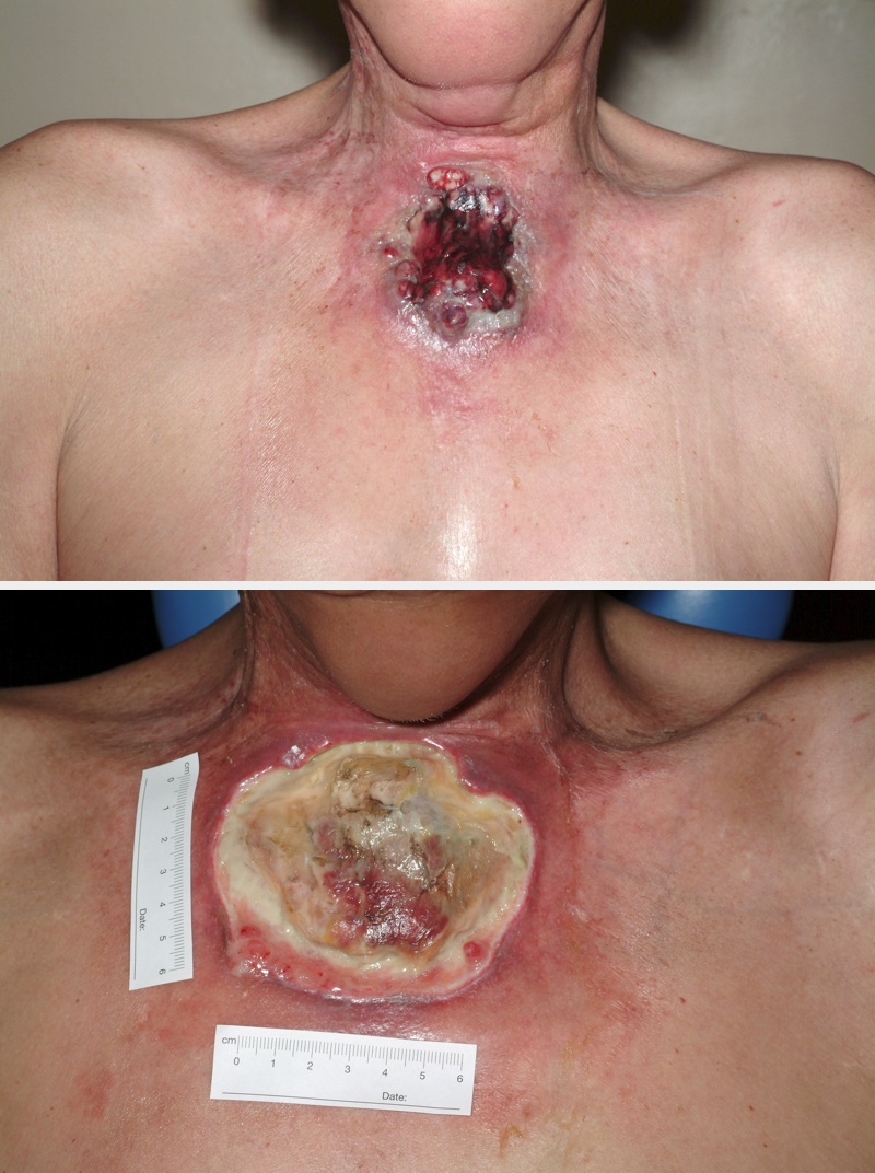
If radiotherapy is the cause of soft tissue necrosis (which may occur together with hard tissue (bone) necrosis and remains a life-long risk after radiotherapy), reconstruction with skin grafts is usually not an option because the blood supply in irradiated soft tissue tends to be impaired. In such cases reconstruction with a flap, carrying its own vascular supply, is the more promising approach. Figure 2 depicts an example of post-radiotherapy necrosis.
Diabetes and other autoimmune conditions, a range of comorbidities, certain medications (such as bisphosphonates), vessel depletion in the neck after neck dissection and the local/regional tissue quality after multiple surgical interventions all considerably increase the risk for the development of soft tissue necrosis. Similar additional risks exist for the development of necrosis of free flaps as well as for the respective donor sites.
When extended and/or deep tissues are affected by necrosis originating from infection, reconstruction of defects after resection may have to be delayed until all infection has been eliminated. The chances of success of surgical reconstruction would be negatively impacted by the continued presence of infected tissues.
Optimal hydration and nutrition, the choice of most suitable antibiotic agents for the treatment of infections, sufficiently radical, possibly repeatedly, resection and suitably timed and planned reconstruction are all crucially important for the successful treatment of soft tissue necrosis (alongside a good portion of patience, perseverance and resilience to frustration).
Treatments of soft tissue necrosis in the maxillofacial region - overview
Oral mucosa
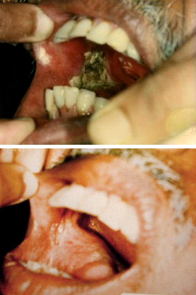
The most suitable treatment protocol depends on cause and extent of the necrosis. Relatively limited necrotic lesions from minor infections or other lesions of the oral mucosa may be treated conservatively. Conservative treatment includes a range of antiseptic mouthwashes, appropriate analgesia and some form of ‘bandaging’ of the oral mucosa by application of a gel that provides a protective thin film as well as some degree of pain relief. Figure 3 shows an example of necrosis of the oral mucosa shortly after photodynamic therapy and after three months.
Where conservative treatment is insufficient and extensive surgical resection is necessary, for reconstruction flaps are always preferable over grafts.
Particularly severe cases of necrosis of the oral mucosa acutely caused by chemotherapy, photodynamic therapy or radiotherapy may be treatment-limiting for these modalities: the treatment may have to be stopped in order to be able to control the mucosal necrosis.
The management options for necrosis of the oral mucosa (and/or skin; see below) from long-term adverse effects of these treatment modalities, in particular radiotherapy are generally similar to the options for acute conditions. Treatment details and options for chronic such necrosis conditions depend on the involvement, or lack of involvement, of hard tissues, especially the mandible.
Skin
Similar to necrosis of the oral mucosa, also for skin necrosis the optimal treatment protocol is dictated by the cause and extent of the necrosis. Minor necrotic lesions from infection or trauma may be treated with the various non-surgical débridement options as appropriate, including topical and/or systemic antibiotic agents as required.
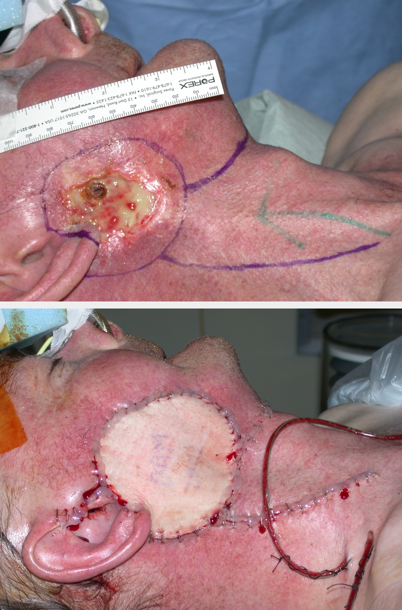
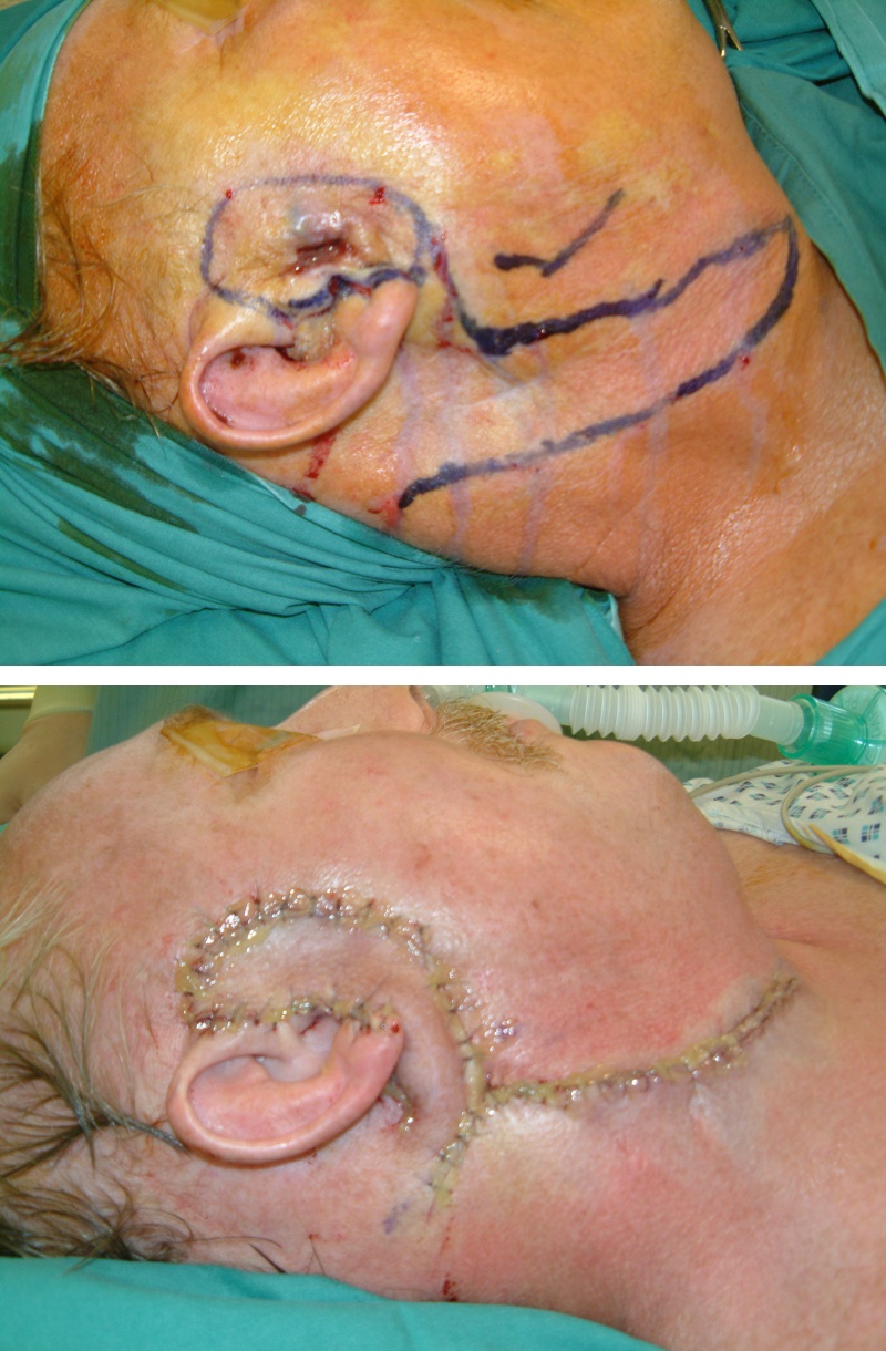
When surgical débridement or more radical resection is the method of choice, resulting relatively minor skin defects in need of reconstruction can be repaired with a skin graft as long as any infection has been cleared. For larger skin defects, like for oral mucosa, reconstruction by a flap is the preferred method. This is especially the case for extended skin necrosis following radiotherapy (Figure 4).
Deeper tissue layers (fascia, muscle)
Necrosis affecting deeper tissue layers, including muscle and fascia, is most likely to be part of a serious infection or may result as a long-term effect from radiotherapy. As such, the most likely treatment options will be (emergency) radical surgical intervention and systemic antibacterial medication when infection is involved (similar in spirits to the surgical management of severe abscesses). These conditions are likely to require radical resection of necrotic tissues, with reconstruction to follow (either immediately following resection (see Figure 2 for an example), or after a delay until eradication of infection has been achieved). Necrosis of deeper tissue layers, such as necrotising fasciitis, may invade bigger vessels and thus carries a risk of serious haemorrhage that may require emergency surgical intervention.